Non Invasive Live Cell Imaging
Acquire real time quantitative imaging for live cells, assays, tissues & organoids.
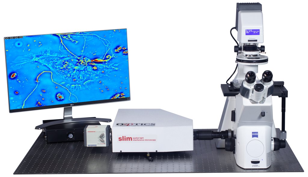
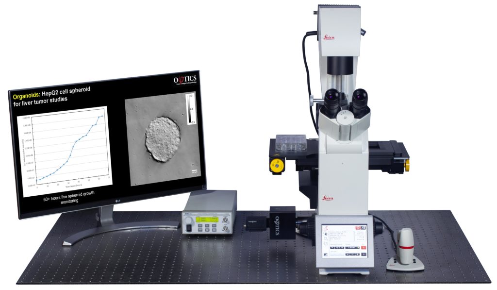



High contrast imaging thin specimens
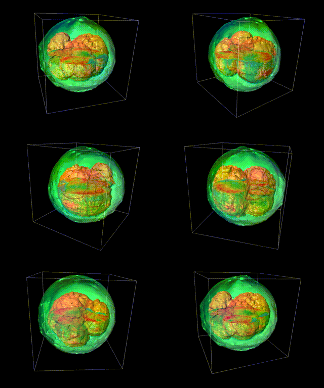
High contrast imaging thick specimens
Deep learning is applied to QPI data

Plugs into your existing Olympus, Lieca, Nikon or Zeiss microscope.
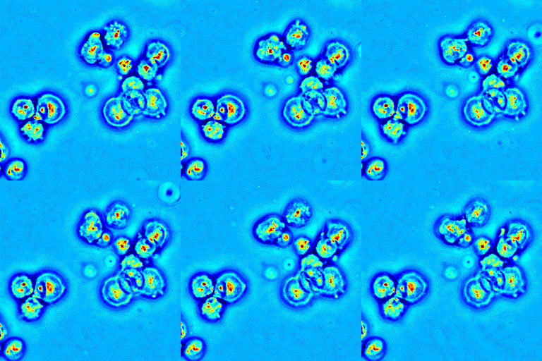
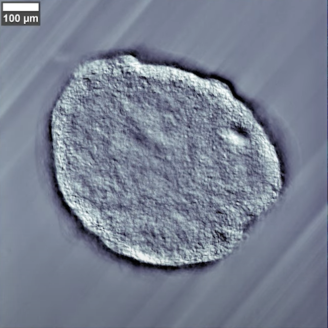
Benefits of QPI for Live Cell Microscopy
Quantitative Phase Imaging refers to a number of techniques to provide contrast by quantifying changes in the phase shift as light propagates a specimen. Phi Optics has invented techniques that provide excellent contrast, with non invasive, label free mapping.
Recent Publications
Cell Cycle Stage Classification Using Phase Imaging with Computational Specificity
Y. He, S. He, M. Kandel, Y. J. Lee, C. Hu, N. Sobh, M. Anastasio, and G. Popescu
ACS Photonics 2022
Cell Cycle Stage Classification Using Phase Imaging with Computational Specificity
Y. He, S. He, M. Kandel, Y. J. Lee, C. Hu, N. Sobh, M. Anastasio, and G. Popescu
ACS Photonics 2022
Live-dead Assay on Unlabeled Cells Using Phase Imaging with Computational Specificity
C. Hu, S. He, Y. Lee, Y. He, E. Kong, H. Li, M. Anastasio, and G. Popescu
Nature Communications, (2022) 13:713
Synthetic Aperture Interference Light (SAIL) Microscopy for High-Throughput Label-Free Imaging
C. Hu, M. Kandel, Y. Lee and G. Popescu
Applied Physics Letters, 119, 233701 (2021)