
Non Invasive Live Cell Imaging
Acquire info in real time with non invasive quantitative imaging for live cells, assays, tissues and organoids



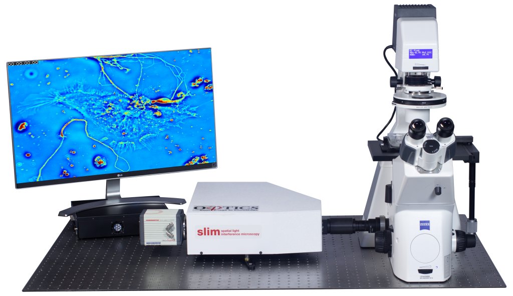
High contrast imaging thin specimens
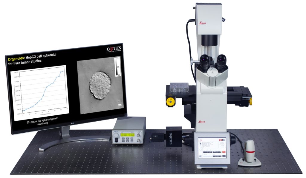
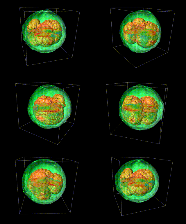
High contrast imaging thick specimens
Deep learning is applied to QPI data
Benefits of QPI for Live Cell Microscopy
Quantitative Phase Imaging refers to techniques to provide contrast by quantifying changes in the phase shift when light propagates through a specimen. Phi Optics has invented techniques that provide excellent contrast, with label-free mapping.
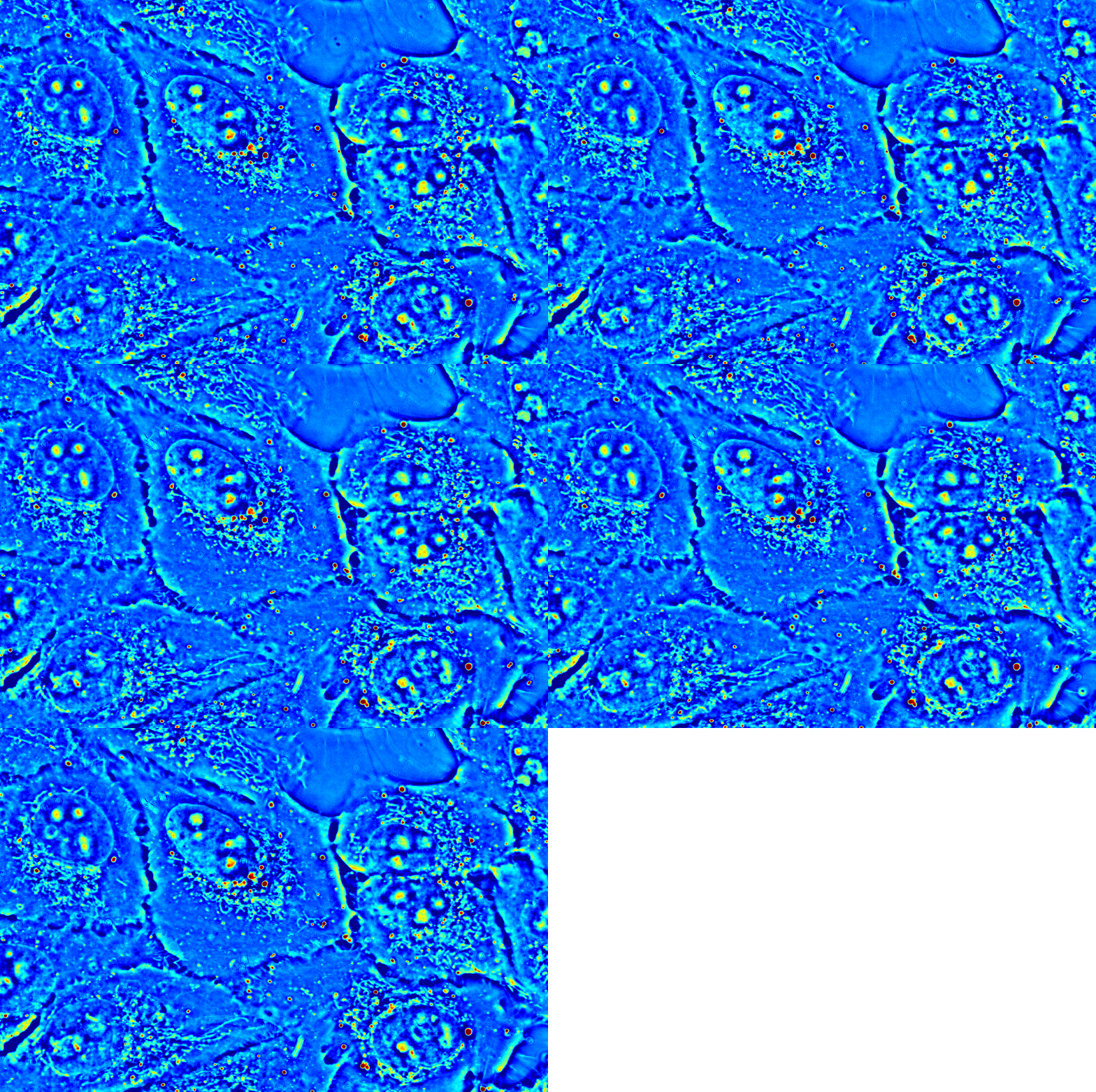
There are important considerations in live cell microscopy that we have addressed.

Phi Optics CellVista acquisition software seamlessly controls the frame motorization of your existing microscope for programmed 4D (3d + time lapse) multichannel (QPI and Fluorescence) monitoring of live or fixed cultures.
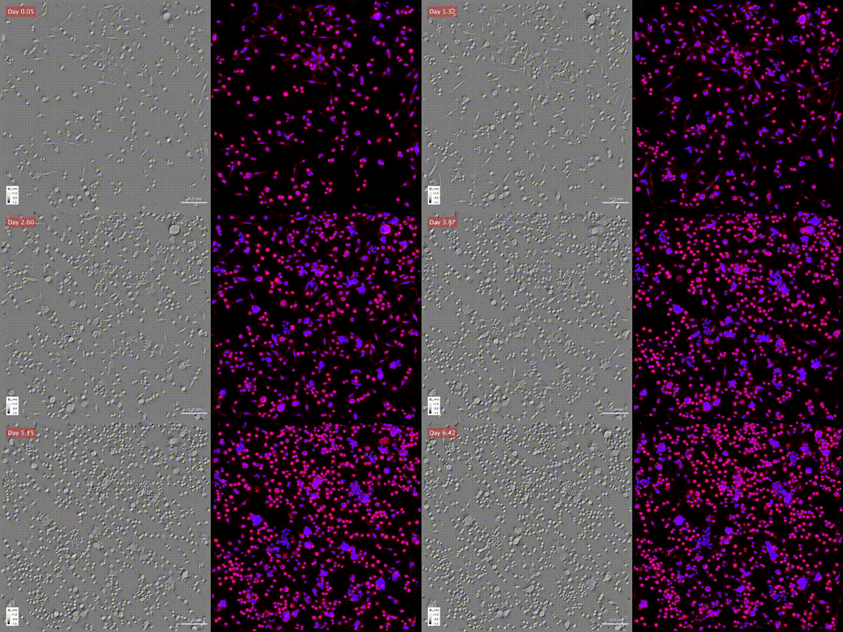
CellVista SLIM is made for optically thin specimens, and CellVista GLIM handles thick specimens, such as embryos, organoids and tissues.
Scientific Articles
2022
Cell Cycle Stage Classification Using Phase Imaging with Computational Specificity
Live-Dead Assay on Unlabeled Cells Using Phase Imaging with Computational Specificity
2021
Synthetic Aperture Interference Light (SAIL) Microscopy for High-Throughput Label-Free Imaging